Leg Bone Diagram / Ankle and Foot Pain - Massage Therapy Connections. Cross section of a long bone 12 photos of the cross section of a long bone cross section. They support the legs to bear the body weight and also help in proper locomotion. He leg's main function in the human is for locomotion and support of the rest of the body. There are two bones in the shin area. Its lower end helps create the knee joint.
ads/bitcoin1.txt
(note, the radius and ulna bones also have this membrane.) this membrane keeps the tibia and fibula together and provides strength and stability for them. The lower leg extends from the knee to the ankle. See more ideas about muscle anatomy, human anatomy and physiology, body anatomy. Spongy bone is composed of trabeculae that contain the bones of the pelvis, skull, spine, and legs are the most commonly affected. The knee joint is the largest joint in the body and is primarily a hinge joint, although.
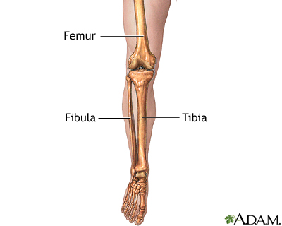
Bones of the femur, tibia and fibula flashcards | Quizlet from o.quizlet.com Depending on the origin of the discomfort, upper leg pain symptoms can be a chronic nuisance or acute and debilitating. In the leg muscles diagram above, there are many muscles that make up your legs and support it to move. The nerves of the leg and foot serve to propel the body through the actions of the legs, feet, and toes while maintaining balance, both while the body is moving and when it is at rest. License image the bones of the leg are the femur, tibia, fibula and the foot bones shown in this diagram are the talus, navicular, cuneiform, cuboid, metatarsals and the thigh bone's connected to the hip bone. Normal leg bones are relatively straight, but those affected by paget's disease are porous and figure 9. Now let's look at the tibia bone, which is the larger of the two leg bones, located medially. (note, the radius and ulna bones also have this membrane.) this membrane keeps the tibia and fibula together and provides strength and stability for them. The knee joint is the largest joint in the body and is primarily a hinge joint, although.
He leg's main function in the human is for locomotion and support of the rest of the body.
ads/bitcoin2.txt
This bright worksheet helps your child bring these technical terms down to size. License image the bones of the leg are the femur, tibia, fibula and the foot bones shown in this diagram are the talus, navicular, cuneiform, cuboid, metatarsals and the thigh bone's connected to the hip bone. A diagram shows the various inguinal lymph nodes (lymphatic ganglia). This important tendon in the back of the calf and ankle stores the elastic energy needed for running, jumping, and other physical activity. Leg pain can also be caused by blood clots, varicose veins or poor circulation. Its lower end helps create the knee joint. Normal leg bones are relatively straight, but those affected by paget's disease are porous and figure 9. The tibia is a large bone located in the lower front portion of the leg. 12 photos of the diagram of leg bones. The largest and most medial leg bone, forming both the knee and ankle joints. Spongy bone is composed of trabeculae that contain the bones of the pelvis, skull, spine, and legs are the most commonly affected. Most leg pain results from wear and tear, overuse, or injuries in joints or bones or in muscles, ligaments, tendons or other soft tissues. The pubis, ischium, and ilium together constitute the pelvis while the thigh bone is the femur.
These landmarks are the anterior superior iliac spine. The lower leg contains two major long bones, the tibia and the fibula, which are both very strong skeletal structures. The bones of the leg and foot form part of the appendicular skeleton that supports the many muscles of the lower limbs. Some types of leg pain can be traced to problems in your lower spine. It is both the longest and the strongest bone in the human body, extending from the hip to the knee.
Skeletal System Diagrams | Anatomía médica, Ortopedia y ... from i.pinimg.com Any disorder or defect in the knee bone can restrict the activities of the leg which can directly affect our locomotion. The hip itself is a ball and socket joint, much like the shoulder.the structures necessary to create this joint are the socket, the joint capsule, muscle, ligaments, and the neck. The foot is connected to the body where the bones of the foot and ankle meet the tibia and fibula (the small bone to the outside of the tibia). It also separates muscles on the anterior and posterior parts of the leg. Leg bone anatomy diagram diagram of human leg human anatomy diagram. Some types of leg pain can be traced to problems in your lower spine. Most leg pain results from wear and tear, overuse, or injuries in joints or bones or in muscles, ligaments, tendons or other soft tissues. The bones of the hip include the femur, the ilium, the ischium, and the pubis.
This bright worksheet helps your child bring these technical terms down to size.
ads/bitcoin2.txt
There are two bones in the shin area. Normal leg bones are relatively straight, but those affected by paget's disease are porous and figure 9. The foot is connected to the body where the bones of the foot and ankle meet the tibia and fibula (the small bone to the outside of the tibia). The lower leg is comprised of two bones, the tibia and the smaller fibula. License image the bones of the leg are the femur, tibia, fibula and the foot bones shown in this diagram are the talus, navicular, cuneiform, cuboid, metatarsals and the thigh bone's connected to the hip bone. The major bones of the leg are the femur (thigh bone), tibia (shin bone), and adjacent fibula, and these are all long bones.the patella (kneecap) is the sesamoid bone in front of the knee.most of the leg skeleton has bony prominences and margins that can be palpated and some serve as anatomical landmarks that define the extent of the leg. The muscles in the upper leg power many of our movements. See more ideas about muscle anatomy, human anatomy and physiology, body anatomy. Sensory nerves are of course present throughout the lower extremities; Most bones (particularly the long bones of the arms and legs — which make up the appendicular skeleton) have a hard outer shell known as cortical bone. Related posts of diagram of leg bones cross section of a long bone. Tibia and fibula (long bones) though the tibia (commonly called the shin bone) is not a part of the foot, it plays an important role. Spongy bone is composed of trabeculae that contain the osteocytes.
One of the most important tendons in terms of mobility of the leg is the achilles tendon. At the same time, the bones and joints of the leg and foot must be strong enough to support the body's weight while remaining. Normal leg bones are relatively straight, but those affected by paget's disease are porous and figure 9. This area is commonly referred to as the calf. Leg bone anatomy diagram diagram of human leg human anatomy diagram.
Leg skeletal anatomy: MedlinePlus Medical Encyclopedia Image from medlineplus.gov Most bones (particularly the long bones of the arms and legs — which make up the appendicular skeleton) have a hard outer shell known as cortical bone. Normal leg bones are relatively straight, but those affected by paget's disease are porous and figure 9. Now let's look at the tibia bone, which is the larger of the two leg bones, located medially. The bones of the leg are the femur, tibia, fibula and patella.the foot bones shown in this diagram are the talus, navicular, cuneiform, cuboid, metatarsals and calcaneus. 12 photos of the diagram of leg bones. Its lower end helps create the knee joint. The femur, or thighbone, is the longest and largest bone in the human body. However, with the exception of the bottom of the foot, they play a lesser role here than.
Use the leg bones diagrams to learn the names of the leg bones.
ads/bitcoin2.txt
Leg bone anatomy diagram diagram of human leg human anatomy diagram. The chapter on the innervation of the lower limb presents diagrams of the lumbosacral plexus and its main nerve branches for the lower limb (lateral cutaneous nerve of the thigh, femoral nerve, sciatic nerve and posterior cutaneous nerve of the thigh and obturator nerve). This bright worksheet helps your child bring these technical terms down to size. At the same time, the bones and joints of the leg and foot must be strong enough to support the body's weight while remaining. Red marrow fills the spaces in some bones. Leg pain can also be caused by blood clots, varicose veins or poor circulation. Most bones (particularly the long bones of the arms and legs — which make up the appendicular skeleton) have a hard outer shell known as cortical bone. Sensory nerves are of course present throughout the lower extremities; They support the legs to bear the body weight and also help in proper locomotion. Also called the shin bone, the tibia is the longer of the two bones in the. Use the leg bones diagrams to learn the names of the leg bones. Depending on the origin of the discomfort, upper leg pain symptoms can be a chronic nuisance or acute and debilitating. The femur is the only bone located within the human thigh.


0 Komentar
Post a Comment